What Is A Total Ankle Replacement Surgery?

By: Allison Benson, Physical Therapy student at Hunter College, graduating in May 2018
Worked with Kristin Romeo, PT, DPT

With injury and with age, the joints of your body can be damaged by osteoarthritis, causing painful, aching joints. This pain can follow you throughout the day. You may feel stiff waking up, feel a dull ache when taking your dog for a walk, or feel a painful grinding as you stand up from sitting or as you climb the stairs.

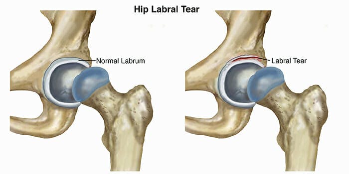
Osteoarthritis (OA) is the most common joint disorder in the United States and is more common in women than men, according to an article published by Zhang & Jordan (2010). In healthy joints, where two or more bones meet and rub together, the bone surfaces are covered by a slippery substance called hyaline cartilage. This cartilage helps make your joints move smoothly and painlessly. With OA, this cartilage has broken down, leaving the bones exposed to each other, creating a grating or “bone on bone” feeling.

When a joint with OA becomes very painful, surgeons often recommend a total joint replacement—you probably know someone who has had a total knee or total hip replacement due to OA. Hips and knees are common sites for OA to develop, both because they move a lot, and because they carry the weight of the body.

You may not have heard of a total ankle replacement, though. Although ankles are also weight-bearing and mobile, they develop OA much less common; only about 1% of the population develops ankle OA (Valderrabano et al., 2009). This means many fewer people have ankle surgeries related to OA.

Another reason you may not have heard about ankle replacement is that it was a relatively unpopular surgery until recently. Total ankle replacements are complicated because there are a lot of important structures packed into a small area at the ankle. They also were associated with a very high failure rate, with surgeons needing to go back in and complete additional surgeries to replace, remove, or adjust the hardware they had placed.

That said, the popularity and success rate of total ankle replacements are on the rise.

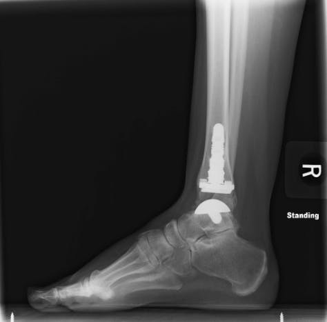
In this surgery, a round metal ball is implanted into the talus, which is an important bone in your ankle. A metal implant is also implanted into the bottom of your tibia, which is the big bone in your calf. A plastic spacer is placed between these two pieces, which allows the tibia to slide smoothly on the talus, just like it does in healthy ankles.

After surgery, a patient will typically be in a surgical boot for 8-10 weeks, and cannot put weight on that foot for 4-6 weeks (Devries, Scharer, & Sigl, 2015). Patients may be referred to physical therapy prior to, or immediately following the procedure for prehab or rehab of the ankle.

Immediately after the surgery, therapists help with gentle work to reduce swelling and pain and prevent tissues from binding down as scar tissue forms. As time passes, therapists help patients regain their strength and range of motion, restoring their ankle to full use. Healing from an ankle surgery is a long process, and requires months of physical therapy, but can be a good option when faced with debilitating ankle OA.

Did you recently have a Total Ankle Replacement surgery? We can help. Schedule your appointment today.

Fill out my online form.


SOURCES

Devries, Scharer, & Sigl. (2015). Total Ankle Arthroplasty Rehab Protocol. BayCare Clinic; Foot & Ankle Center.

Zhang, Y., & Jordan, J. M. (2010). Epidemiology of Osteoarthritis. Clinics in Geriatric Medicine, 26(3), 355–369. https://doi.org/10.1016/j.cger.2010.03.001. Retrieved from
https://www.ncbi.nlm.nih.gov/pmc/articles/PMC2920533/

Valderrabano, V., Horisberger, M., Russell, I., Dougall, H., & Hintermann, B. (2009). Etiology of Ankle Osteoarthritis. Clinical Orthopaedics and Related Research, 467(7), 1800–1806. https://doi.org/10.1007/s11999-008-0543-6

Spinal Stabilization Exercises and Their Role in Alleviating Lower Back Pain

By Boris Gilzon, PT, DPT, OCS, CHT

The Effectiveness of Spinal Stabilization Exercises for Back & Neck Pain

There is no standard approach treating chronic lower back and neck pain. Although this may be unfortunate for many patients to hear, the good news is that there are many conservative methods to alleviate pain.

While conditions like degenerative disk disease, spondylolesthesis, lumbar and cervical radiculopathy are rarely cured completely by conservative measures alone, physical therapy does offer a fair amount of pain relief in the long run.

By utilizing spine stabilization exercises, our physical therapists are able to help patients reduce back and neck pain. This is an active form of treatment requiring the patient to perform exercises to strengthen the muscles and improve the stability of the spine.

Igor assisting his patient with a spine stabilization exercise.

Patients suffering from chronic spinal pain should be leery of physical therapists who mainly offer passive modalities. Examples of passive modalities include heat, electrical stimulation, and massage. Patients should be aware that passive therapeutic modalities do not have sufficient evidence to support their use in chronic spinal conditions.

Spinal stabilization exercises offer the empowerment of the patient and have plenty of research and evidence to support their effectiveness.

Pilates offers an excellent variety of spine stabilization exercise.

Extensive benefits in treating the spine of those who suffer from lower back pain have been discussed extensively in medical literature. Physical Therapists specializing in the spinal disorders are trained in recognizing the factors that affect spinal stability.

Igor Kozlov, DPT treating patient using TRX for back exercise

Components Affecting Spinal Stability

The concept of spinal stability is relatively new with research beginning during the 1970’s.

There are three components that affect spinal stability.

The first component is the passive spinal element: the bone and ligamentous structures. Studies of the cadaver spine in which the bones and ligaments are intact but the muscles were removed showed to buckle under about 20 pounds.

Spinal Ligaments - Medical Illustration Originally Sourced from Kenhub.com
Ligaments of the thoracic spine: posterior (a), anterior (b), lateral (c) and posterior with vertebral arch removed (d). 1, anterior longitudinal ligament; 2, posterior longitudinal ligament; 3, intervertebral disc; 4, ligamentum flavum; 5, intertransverse ligament; 6, supra- and interspinal ligament; 7, radiate ligament; 8, costotransverse ligament.
Originally sourced from: https://musculoskeletalkey.com/anatomy-of-the-thorax-and-abdomen/

The second component of spinal stability are the muscles that surround the spine. The muscular component provides a necessary ‘stiffening” of the spinal segment. In a healthy spine, a very modest level of muscular activity can create a sufficiently stable joint. In a degenerative disk disease, for example, there is more demand on the surrounding musculature. More strength and endurance reserve is needed to overcome an injury and pain.

Deep Muscles of the Back
Deep Muscles of the Back. Medical Illustration originally sourced from: https://pulpbits.net/7-deep-muscles-of-back-anatomy/the-deeper-muscles-of-the-back/

The third component of spinal stability are the neural elements: the central nervous system and peripheral nerves. They are akin to an orchestra conductor, coordinating the performance of various muscles, making sure they are firing at the right time, at the right amount of force.

Spinal Cord Nerves Originally Sourced from Health Jade
Spinal Cord and Nerves – Medical Illustrations originally sourced from https://healthjade.com/spinal-cord/

Multiple studies have shown patients with lower back pain make a “repositioning error” in which their spine would resume to its original position causing pain after performing a certain movement more than patients with a healthy, stabilized spine.

In physical therapy language, we call it a poor postural control.

Specific physical therapy exercises and treatment has shown effectiveness in treating chronic spinal pain.

Lumbar stabilization exercises improve muscular function which can, in turn, compensate for the structural damage to the spinal segment. A thorough dynamic assessment of the spine helps identify postural deficits.

A thoughtful exercise program is designed for each individual by the physical therapist based on their initial testing and evaluation. The most tangible benefit of a lumbar stabilization is that it gives a patient the tools to control their pain.

Interventional Pain Management

Going beyond the scope of physical therapy, interventional pain management is another passive option for chronic spinal pain. This approach serves as a temporary source of relief for patients dealing with low or medium levels of lower back pain. These techniques include performing procedures directly at the level of your dysfunction.

A pain management physician gains access to the areas causing lower back or neck pain by penetrating the surface of the skin. There is a plethora of interventional pain management options for the diagnosis and treatment of the spinal pain.

Epidural steroids are the most common example of the interventional spine management. However, the accuracy and effectiveness of interventional methods in managing lower back pain are not always clear.

In the comprehensive review article published in Pain Physician, 2013 Apr:16, the authors conducted a systematic review of literature in order to collect evidence for the effectiveness of various interventional pain management techniques in the treatment of chronic spinal pain.

The author came to the conclusion that the evidence was fair to good in 52% of therapeutic interventions. The evidence for diagnostic value fared slightly better at 62%.

One significant drawback of all passive techniques is that they do not require a participation of the patient. Without an active engagement of the patient, there is a limited self-control and independence in managing their own condition.

Do you suffer from chronic neck or back pain? Our therapists can help. Schedule your appointment today.

Fill out my online form.

What is the Schroth Method?

Written by Kristin Romeo, DPT
Edited by Alex Ariza

What is Scoliosis?

curvature scoliosis diagram

Scoliosis is a three-dimensional abnormal curvature of the spine. Everyone’s spine has a natural curvature to it, however, if that curvature progresses beyond a certain degree it can be classified as scoliosis. Scoliosis occurs equally among genders but girls seem to be more likely to have scoliosis that has progressed to a level that requires treatment. There are an array of health issues that can accompany scoliosis such as abnormal breathing patterns, visible prominences and poor posture due to muscular imbalances.

What causes Scoliosis?

Scoliosis X-Ray
X-ray image of a person with Scoliosis. Original Image Source: https://www.orthobullets.com/spine/2053/adolescent-idiopathic-scoliosis

There are several different types of scoliosis, however, the majority of scoliosis cases are idiopathic, meaning it has an unknown origin. Idiopathic scoliosis typically begins at a young age and becomes more pronounced during periods of rapid growth.

What are the symptoms of scoliosis?

Pain does not always accompany scoliosis. Scoliosis can present in a variety ways such as abnormal trunk lean, uneven rib cage/shoulders or even back pain. If you suspect scoliosis contact your primary care provider to address your concerns. Prior to Schroth treatment, an x-ray is needed as scoliosis can present differently externally due to overlying musculature and does not give us the full picture. Knowing the bony anatomy allows us to monitor your progress and tailor your treatment to your specific curvature.

rib cage scoliosis cross section diagram

What is the Schroth Method?

Kristin Romeo, DPT Treating Patient with Schroth Exercise 5 | Park Sports Physical Therapy

The Schroth Method is a conservative form of scoliosis treatment designed to target the flexible, postural component of scoliosis. The method was created in Germany in the 1920’s by Katharina Schroth as a way to treat her own scoliosis. Since then the method has made its way across the globe, only recently in the US. Scoliosis specific exercises are targeted specifically to each patient’s curvature through the use of five principles of correction. Subtle postural corrections, spinal distraction and isometric tension help to increase muscle activation and strength in a neutral spinal alignment.

What can I expect during a session of Schroth method physical therapy?

On your first visit, you will be fully evaluated. We will take a look at your posture, your muscular imbalances, address any goals or concerns you may have and take a variety of measurements. You will be sent home the first day with the beginnings of a home exercise program. During your follow-up treatment sessions, we will be utilizing a variety of equipment such as a Schroth wall ladder, physioball, rice bags and Therabands. You will learn about the five principles of correction (1. Auto-elongation (detorsion); 2. Deflection; 3. Derotation; 4. Rotational breathing; and 5. Stabilization) and how to implement them into your home exercise program. We will discuss safe ways to lift, sit and postural corrections to integrate into your daily routine.

Do you or your child suffer from Scoliosis or Kyphosis? We can help. Schedule your evaluation today.

Fill out my online form.

Shoulder Impingement Syndrome Treatment

Written by Nicole Liquori, DPT
Edited by Alex Ariza

In this article, we take a look at the process of one of our sports rehabilitation therapists, Nicole Liquori, DPT. From the initial evaluation to the treatment plan to the patient’s progress throughout, we will get to see and understand the physical therapist’s perspective. On the flip side, we’ll also get to see the patient’s point of view.

Conrad arrived at our facility with complaints of pain and loss of range of motion in his right shoulder. In early 2017, Conrad had been in a swimming accident which left him with transient paralysis. He regained full function of his arms and legs within a few weeks of the accident but was left with the residual weakness of both upper and lower extremities.

Upon evaluation, Conrad demonstrated signs and symptoms consistent with a diagnosis of shoulder impingement syndrome. Conrad presented with rounded shoulders and weakness of his postural and rotator cuff musculature which can strongly affect the mechanical relationship of all joints associated with shoulder mobility.

Conrad’s symptoms included pain and restricted motion when lifting his arm above head and reaching behind his back.

Our treatment initially focused on restoring his normal shoulder and scapular range of motion (as compared to his left shoulder) using mobilization, soft tissue work and passive/active assistive range of motion. Once we were able to establish the normal glenohumeral rhythm – the coordinated motion of the scapula and humerus experienced during shoulder movement – we moved into scapular and rotator cuff strengthening and stabilization activities.

We focused on functional movements that would translate into his activities of daily living (i.e., reaching for a cup in a high cabinet), as well as recreational activities (i.e., throwing a ball, swimming, etc.).

Conrad’s treatment was cut short secondary to surgery on his spine, but prior to discharge, Conrad had returned to performing most everyday tasks, as well as throwing a ball overhand without pain.

In Conrad’s Own Words

I highly recommend Nicole Liquori at Park Sports PT. After a serious accident, I was unable to raise my arm over my head. I couldn’t throw a ball or swim with an overhand stroke. Nicole changed that in the space of two months. A combination of deep massage, passive movement and guided exercises brought back pain-free use of my shoulder. Her knowledge is apparent in her explanations of the functional basis for the exercises. Her skill is demonstrated in her wonderful touch. She confidently employs just the right amount of force in the right places. On top of that, Nicole is sympathetic and encouraging. She has all the qualities of a first-rate therapist and she helped me immensely.

Conrad L.

Do you currently suffer from Shoulder Impingement Syndrome? We can help. Schedule your appointment today.

Fill out my online form.

Couples that do Physical Therapy Together…

Married couple, Michael and Lila R., are longtime patients of Park Sports Physical Therapy. Michael is a lifelong athlete having run 26 marathons during his lifetime. He has also been a member of the Prospect Park Track Club, a local running club in Park Slope, Brooklyn. Michael has over 30 years of running under his belt. That’s a whole lot of miles!

His wife Lila, also an athlete, has spent most of her life swimming and running.

Lila’s first experience at Park Sports started back in 2011. She came in with a rotator cuff condition. One of our therapists treated her and got her back to swimming fairly quickly. Lila recommended her husband, Michael also get treated at Park Sports after her positive experience.

Michael suffered from arthritis in both his knees. It was when he tripped and injured his iliotibial band that he came in for treatment. He was feeling a snapping sensation whenever he would walk up and down stairs.

Kristin Romeo, DPT became both Michael’s and Lila’s therapist, often times seeing them at the same time for treatment. For Michael, Kristin used manual therapy, stretching, strengthening exercises, and worked on improving his balance on the injured leg.

During the interview, Michael and Lila spoke very highly of Kristin, saying “Kristin is attentive, listens to your needs, and makes sure that the problem area is getting the right treatment.”

“The sessions hold me accountable,” Michael mentioned. The exercises have helped him greatly throughout his treatment. He’s progressed to the point where he no longer feels any snapping sensation walking down the stairs.

Lila similarly was very pleased with her progress.

Michael and Lila, being older in age, both realize the importance of maintaining balance to avoid falls. Physical therapy has proven to be quite useful in that regard.

Both Michael and Lila recommend the services at Park Sports highly saying, “almost all our friends in the neighborhood go to Park Sports for physical therapy. This is a great place that offers excellent care. The therapists that work here are excellent. We couldn’t be happier with Kristin’s care.”

Frozen Shoulder Treatment – Regaining Mobility Through Physical Therapy

Written by Edward Umheiser, PT, DPT
Edited by Alex Ariza

Injuries to the shoulder are fairly common in everyday life. Strains and sprains resulting from repetitive activities such as throwing a ball or twisting your arm while reaching behind your car seat happen all the time, and usually heal on their own in only a few days. But what happens if your shoulder suddenly starts to lose its normal range of motion for an unexplained reason?

In this article, I would like to talk about the diagnosis known as adhesive capsulitis, more commonly referred to as frozen shoulder.

What is Frozen Shoulder?

Frozen shoulder is a fairly common diagnosis, but is not well understood and does not always have a known cause. Many people develop frozen shoulder following a surgical procedure to the shoulder, or after an injury that results in the shoulder needing to be immobilized for a short period of time. However, in some cases, some people develop symptoms with no known trigger.

The symptoms are fairly clear-cut – a marked loss of normal range of motion and stiffness of the shoulder joint that may or may not be accompanied by pain.

This shoulder stiffness affects motion in all directions such as reaching up in the air or trying to place the hand behind the back or behind the head. Adhesive capsulitis begins as an inflammatory process within the shoulder and leads to scar tissue formation which can restrict shoulder motion.

For some people, this scar tissue formation can lead to pain in the shoulder joint, especially with movement and during the evening hours while trying to sleep.

The Stages of Frozen Shoulder

Physical Therapist, Edward Umheiser, DPT treating patient with frozen shoulder.
Physical Therapist, Edward Umheiser, DPT treating a patient with frozen shoulder.

There are several stages of frozen shoulder each with their own unique characteristics:

Inflammatory Stage

The inflammatory stage lasts approximately three months and is characterized by pain with shoulder movement in most directions. Pain is often sharp with movement at the end of the range, and there is an ache at rest. Pain is also commonly felt at night, making it difficult to fall asleep.  The range of motion may still be normal at this early stage.

Freezing Stage

Edward Umheiser, DPT measures the shoulder's range of motion.
Edward Umheiser, DPT measures the shoulder’s range of motion.

The freezing stage typically lasts 3-9 months. This is when flexibility of the shoulder begins to reduce due to pain. At this time, people often begin to notice difficulty reaching overhead, or reaching behind their back. This stage, known as the frozen stage, is when the stiffness is most severe. The inflammatory process inside the shoulder joint is starting to decrease at this stage so pain is lessened or non-existent. Over the past several months a thick layer of scar tissue has formed around the shoulder capsule, which makes it difficult to move. This is the stage when most people are diagnosed with frozen shoulder.

Thawing Stage

Edward Umheiser, DPT treating a patient with frozen shoulder. With consistent physical therapy sessions, a patient can begin seeing an increased range of motion.

Finally, the thawing stage is when shoulder motion gradually starts to return to normal. This stage can last anywhere from 9 months to two years and may leave some minor limitations in shoulder range of motion afterward.

Treating A Frozen Shoulder

Edward Umheiser, DPT teaching the patient shoulder exercises.

Physical therapy along with treatment provided by your doctor such as cortisone injections are the first line of defense in reducing the time a patient may experience the symptoms related to a frozen shoulder.

A doctor will typically take some images of the shoulder to rule out other possible structural involvement such as the rotator cuff or the labrum.

At that point, physical therapy treatment 2-3x a week is generally recommended where manual shoulder stretching, massage, mobilization, and exercises are performed to keep the shoulder as pain-free as possible, and to help increase the range of motion.

It is also important to continue to perform the prescribed stretching exercises at home under the guidance of a physical therapist. Generally, the time it takes for a frozen shoulder to “thaw” can be halved under physical therapy treatment. Most patients do not need surgical intervention to correct this diagnosis.

Steps to Take If You Suspect Having a Frozen Shoulder

If you have noticed that you have a lot more difficulty with overhead shoulder motion following a shoulder injury, let your doctor know. If this has persisted for several months, it may be a sign of adhesive capsulitis. An orthopedist can help you make that diagnosis, and physical therapy can help you get onto the road to recovery.

Schedule Your Physical Therapy Appointment Today To Begin Frozen Shoulder Treatment

Fill out my online form.

Treating Hip Pain for Brooklyn Athletes

Written by Boris Gilzon, PT, DPT, OCS, CHT & Alex Ariza

Physical Therapists play an essential role in the nonsurgical treatment of hip pain. Two of the most common conditions associated with hip pain are labral tears and hip impingement.

Labral Tears

Labral tears occur when the labrum, which is the cartilage that comes between the head of the femur – your thigh bone – and your pelvis – your hip bone, begins to wear down or experiences trauma. The most common cause of labral tears is repetitive stress on the hip joint, especially for athletes participating in running and skating sports.

Labral Tear - Hip Pain
Original Image Source:
https://www.moveforwardpt.com/symptomsconditionsdetail.aspx?cid=fabdfb4e-5fb5-4077-b341-df5d04a93605

Hip Impingement

Hip impingement, also known as femoroacetabular impingement (FAI), occurs when extra bone grows on either or both the head of the femur or the acetabulum (the part of the pelvis where the femur meets – the “socket”). The extra bone creates an irregular fit and can start to rub against the cartilage or bones in a way that can damage the joint causing hip pain.

There are 2 types of hip impingement. They can occur separately or together.

Hip Impingement Types
Original image source:
https://orthoinfo.aaos.org/en/diseases–conditions/femoroacetabular-impingement/

Cam-type Impingement

The head of the femur is shaped like a sphere. It acts as the “ball” for “ball and socket” joint in your the hips. In this deformity, an excessive bone growth around the head of the femur creates an irregular shape that makes it difficult to rotate in the acetabulum, your “socket”.

The misshapen overgrowth contacts the cartilage that lines the hip socket, the labrum, and can cause it to become worn and frayed. This can lead to hip instability and pain.

Pincer-type Impingement

Pincer impingement is when the acetabulum (hip socket) protrudes around the femur head, decreasing the space of the joint. Overtime with repetitive contact, hip flexion (bending over, sitting, walking) leads to tearing and inflammation of the cartilage that envelopes and protects the joint (labrum).

Symptoms

With either one of these conditions you may experience:

  • Increased pain with prolonged sitting and leaning forward in the sitting position.
  • Sharp pain during squatting, changing direction during running, pivoting and lateral motions./li>
  • Stiffness and deep aching in the front of the hip. Occasional groin pain.
  • Decrease ability to turn your thigh inward.

Treating Hip Pain with Physical Therapy

Kristin Treating Patient - Hip Mobility 2 | Park Sports Physical Therapy

Treatment of hip pain starts with a comprehensive evaluation of movement of your hip and surrounding joints.

Our therapists will test the strength of the muscles around your hip and observe your movements to properly diagnose the condition. During each session, our therapists will instruct and educate you about your body mechanics, the origin of your injury, and exercises you can perform to prevent future injury to the hip and strengthen the muscles around the hip and other parts of your body to stabilize and correct any imbalances.

Your orthopedic doctor may order X-rays and an MRI to confirm the diagnosis.

Hip impingement and labral tears may be concurrent with lower back pain and pelvic floor dysfunction.

Once your weaknesses and imbalances are identified, the goal of therapy is to restore normal force coupling and to develop strategies to compensate if your condition is chronic.

Strengthening of weak muscles and stretching tight muscles can help to avoid the progression of hip impingement. Manual therapy techniques may also be employed restore flexibility of the joint.

At Park Sports Physical Therapy, one of our therapists, Svetlana Lazarev, PT, is an advance hip clinician certified by the Hospital for Special Surgery (HSS).

Hip Arthroscopy Post Operative Rehabilitation

If you do end up having surgery for your hip pain, our therapists follow an established post-surgical protocol. Physical therapy is known to shorten recovery time and ensures that your body is healing properly. Our therapists help to promote safe healing by limiting the weight you put on the operated leg, exercises, stretching, and manual therapy.

The next stage of postoperative rehabilitation focuses on improving your range of motion and strength. During your treatment, our physical therapists can keep your doctor and orthopedic surgeon informed of your progress if you choose.

After 4 to 6 months of treatment following surgery, you’ll be able to return to your normal activities.

Do you currently suffer from hip pain? We can help.

Fill out my online form.

Why Men Should See A Pelvic Floor Therapist

Written by Alex Ariza
Reviewed by Irene Hernandez, PT, DPT

Everyone has pelvic floor muscles. So why is it that almost all information relating to pelvic floor therapy focus solely on women?

There are obvious reasons why Pelvic Floor Therapy is normally associated with women’s health issues. For starters, Pelvic Floor Dysfunction is far more prevalent in women than men, especially pregnant women.

Although this is true, there is still a fair amount of men suffering from symptoms associated with pelvic floor dysfunction. What’s worse is that most of them don’t even know it.

In fact, many times even medical doctors can misdiagnose pelvic floor dysfunction with much more serious conditions and diseases since symptoms can feel very similar to them. Sometimes this can lead to prescribing unnecessary medications and antibiotics, and in more extreme cases, surgery.

All of this could be preventable if men were more informed about their bodies and if medical practitioners knew more about pelvic floor issues.

When men suffer from erectile dysfunction, painful urination, frequent & involuntary urination, painful ejaculation, constipation, or pain after having a bowel movement, the last thing they would suspect is a problem with their pelvic floor muscles. And who can blame them?

Pelvic Floor Therapy for Men

At Park Sports Physical Therapy, part of our mission is to educate our patients about their bodies and the reasons they feel the symptoms that they do. We also stress the importance of preventative care, and the steps they can take through exercise and posture to keep them functional.

Our pelvic floor specialist, Irene Hernandez, DPT, specializes in treating both women AND men. We strongly encourage our male patients suffering from any of the conditions or symptoms listed above to come in for an evaluation.

Learn more about the pelvic floor and the different conditions associated with pelvic floor dysfunction by visiting this page.

The pelvic floor plays a few roles. For one, it is a group of muscles that form the “bottom” of your body’s core. This keeps the organs in place and from dropping out of the pelvis.

The second major responsibility of the pelvic floor is the control of the sphincters. This includes the anus and urethra. Weak pelvic floor muscles can result in incontinence – or the inability to hold in urine and/or feces. An over contracted pelvic floor will make it difficult to make a bowel movement, leading to constipation, or the inability to release urine.

The third major responsibility of the pelvic floor is sexual function. For men, dysfunction of the pelvic floor can lead to erectile dysfunction (ED). There are many other factors that can play into ED, but seeing a pelvic floor specialist can alleviate and correct muscles related to healthy sexual function.

Male Pelvic Floor Medical Illustration - Illustrated by Amy Stein - Heal Pelvic Pain
Male Pelvic Floor Medical Illustration – Illustrated by Amy Stein – Heal Pelvic Pain
Original Source: https://www.healpelvicpain.com/

Schedule Your Pelvic Floor Evaluation Today.

Fill out my online form.

Here’s What You Can Expect For The New Year In Terms Of Your Insurance

Dear Patients,

With the new year approaching, changes to your health insurance policy may occur. These changes may affect your current payment plan.

If you have an insurance plan with deductible policies – please be aware that your accumulations will reset for the new year. This means your deductibles will have to be met again before the health plan starts covering physical therapy sessions.

If you have questions regarding the costs of your physical therapy visits for the new year, please give us a call at (718) 230-1180, and we can give you an estimated dollar amount.

If you are switching to a new insurance – please give us a call with your new policy information. We can reach out to your insurance company to verify your benefits and coverage for your convenience.

If you decide to keep your current insurance provider – please be aware that although your insurance remains the same, coverage may differ for the new year.

Unfortunately, we are not able to verify your benefits with your insurance company until the new year begins. We want to assure you that we are working to ensure you with accurate benefit information.

We strongly encourage and recommend that you call your health insurance provider to obtain benefit information for additional information.

Please call us at (718) 230-1180 or email us at info@parksportspt.com if you have any questions about your insurance.

Best,

Times Fong & the Park Sports Physical Therapy Team

Times Fong
Office Manager
(718) 230-1180

What We Can Learn From Angels Pitcher Shohei Ohtani’s Elbow Injury

Written by Boris Gilzon, PT, DPT, OCS, CHT and Alex Ariza

If you’re a fan of baseball you may have heard that New Los Angeles Angels pitcher, Shohei Ohtani, has a damaged Ulnar Collateral Ligament (UCL) in his pitching arm.

This is bad news for Ohtani, as his career relies heavily on the function his elbow. Although reports show that Ohtani only has a first-degree sprain, consistent use of his elbow can present serious problems down the line.

In the world of physical therapy, elbow pain does not get enough exposure and is often ignored. More often than not, people continue performing their daily activities that can exacerbate injury to the elbow and cause damage to the joint and ligaments.

Ulnar Collateral Ligament (UCL) Injury

As Physical Therapists, we see sports injuries in a different light, especially when they make the news. This post will share some of the intimate knowledge we possess from years of treating a number of injuries and conditions that athletes may face.

The Ulnar Collateral Ligament, located in the elbow, is a complex structure consisting of three bands. It is challenged more when the elbow is in flexion. During full elbow extension, the bony congruity provides additional stability.

Ulnar Collateral Ligament Anatomy

Ulnar Collateral Ligament injury is common in many overhead sports. The forces that can lead to injury on the elbow are generated when the elbow goes from flexion to extension at a high velocity. In Ohtani’s case, this would be frequently pitching at a consistent speed of 100 mph.

This injury occurs from repetitive valgus (a condition in which the bone segment distal to a joint is angled outward) stress on the medial (inner aspect) of the elbow. This kind of elbow sprain commonly occurs in baseball pitchers. It is less frequent in racket sports, volleyball, and hockey, but can still occur in athletes playing those sports.

Damage to the ligament can be caused by sudden trauma or a gradual stress. The most typical sign is a pain in the inner elbow while performing a physical activity. Patients commonly describe an elbow sprain as a twinge or sharp pain when pushing up, such as getting up from the chair.

If a ligament is compromised a patient may develop:

  • A sense of looseness or instability in the elbow.
  • Irritation of the ulnar nerve (aka the “funny bone”): This is felt as a tingling sensation or numbness in the small finger and ring finger.
  • Decreased ability to throw a baseball or other object overhead.

Treatment for Ulnar Collateral Ligament Injury

Ulnar Collateral Ligament injury can happen to anyone at any age. Parents and coaches should be aware of the issue and be alert if the child complains of the pain in the elbow.

Physical Therapy is the best first line of defense following this diagnosis. At Park Sports Physical Therapy, our Physical Therapists are skilled and experienced in treating elbow instability and ulnar collateral ligaments tears.

The treatment approach is based on thorough examination and biomechanical analysis of the activity that caused the condition.

It includes:

  • Strengthening weak muscle group
  • Activity modification.
  • Manual therapy and therapeutic modalities.

Post-Surgical Recovery

In cases of severe damage and instability, the ligament is reconstructed in what is commonly known as a Tommy Johns procedure. Physical Therapy treatment in this case follows an established post-surgical protocol. Our therapists work closely with an Orthopedic Surgeon to optimize post-surgical recovery and achieving the highest functional outcomes.

Do you suffer from Ulnar Collateral Ligament Injury? Take the first step to get out of pain by scheduling your appointment.

Fill out my online form.