The Schroth Method for Scoliosis

 
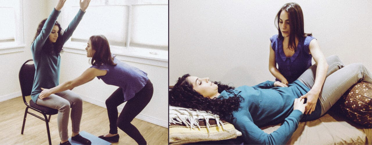
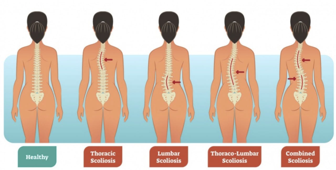
The Schroth Method for scoliosis utilizes a three dimensional approach to elongate the trunk and correct imbalances of the spine. The goal is to develop the inner muscles of the rib cage in order to change the shape of the upper trunk and to correct any spinal abnormalities.

Developed by Katharina Schroth

The Schroth Method was originally developed by Katharina Schroth in Germany. The method uses customized exercises to return the body’s posture to a more natural position. It is standard treatment for scoliosis in many European countries, and is gaining trust and popularity in the United States.

Scoliosis Symptoms

Symptoms of Scoliosis vary. Pain does not always accompany scoliosis. As a result, scoliosis can present in a variety ways such as abnormal trunk lean, uneven rib cage/shoulders or even back pain. If you suspect scoliosis contact your primary care provider. Prior to Schroth treatment, an x-ray is needed as scoliosis can present differently externally due to overlying musculature and does not give us the full picture. So, knowing the bony anatomy allows us to monitor your progress and tailor your treatment to your specific curvature.
Treatment at Park Sports PT

Getting treatment at Park Sports PT means you are working with certified therapists trained in the Schroth Method. They are partners in your health and wellness. Your therapist will create a personalized plan just for you and your specific condition. Our treatment approach can treat scoliosis patients of all ages. It can also can be utilized to treat in all stages of scoliosis, including after surgery.

Throughout treatment, we look closely at the three-dimensional curve in the spine. We teach patients very specific ways to correct that curve or scoliosis posture. Once patients have recognized their corrected posture, we teach them breathing and muscle activation techniques to hold that correction. Essentially, we help train the motor neurons to sense when they’re in a corrected posture versus when they’re in their scoliosis posture.

The Schroth Method takes a lot of repetition and commitment from the patient to learn these techniques. That’s why home exercises are also recommend at least five days a week for a half hour each time.

Trial results show improved patient outcomes

Several trials have found that physical therapy scoliosis-specific exercises lead to improved patient outcomes. This includes less pain and improved muscular strength, muscular endurance and self-image. In one study, spinal deformity improved in 69 percent of patients who completed Schroth exercises. This compared to only 6 percent in patients who did not complete their exercises. The Schroth Method at Park Sports PT works.

Your Park Sports PT team will create a personalized plan and will help prepare you to work on your strength and posture at home. The result is long term care and a stronger and healthier body.

Contact Us

If you or your child has any symptoms or have visited your primary care provider and are looking for treatment, contact us today to learn more. We are here to help.

Fill out my online form.


Pelvic Floor Therapy

Pelvic Floor Therapy and Dysfunction.

Pelvic Floor Therapy can be an important part of a healthy life.  So when it comes to posture, comfort, intimacy, and wellness, a healthy pelvic floor is one of the most vital parts of your body. Many women face daily discomfort, lack of intimacy, physical pain, and more due to a dysfunction of the muscles, ligaments, and nerves that support the pelvic region.

A healthy and re-energized pelvic region can greatly restore your well-being and help to eliminate symptoms that are negatively effecting your life.

Pelvic Floor Dysfunction (PFD) may be brought on by a variety of reasons. These include pregnancy, childbirth, menopause, prior surgery or radiation, heavy lifting and straining, obesity, and congenital weakness. This can lead to vulvar pain, localized pelvic discomfort, prolapse, low back pain, leakage, incontinence and much more. Most do not seek advice from a physical therapist. Those who do however, can see life changing results.

At Park Sports, we are dedicated to life changing results.

Park Sports Therapy Approach to Pelvic Floor Dysfunction.

What to Expect When Getting Started.

At Park Sports, we understand the seriousness of your decision to seek help. We offer evidence-based and individualized treatment plans backed by a wealth of knowledge and a history of success.

Our pelvic floor therapist Lacey Salberg PT, DPT has received extensive training from the Herman & Wallace Pelvic Rehabilitation Institute, a leader in pelvic floor education and awareness. Lacey will take the time to discuss your health concerns. She will also fully evaluate your condition and create personalized plan, uniquely designed for your comfort level, your lifestyle and your body. You will receive one-on-one sessions in our private treatment room as she coaches you towards your health goals.

Working Together.

We consider your health a team effort. Part of your therapy will include an at home exercise program that will help you progress through your treatment plan. As you make advancements, it is possible that your initial plan may need some adjustments. Your therapist will listen carefully to your body and closely monitor your functional gains. If necessary, she will discuss modifying your treatment plan to better achieve your health goals.

Long-Term Planning.

Once you reach your goals, your therapist will provide an at home plan. This plan will help you maintain your new functional gains and pelvic floor health. Of course we will always be available to provide advice and assistance. As your health and wellness partner – our goal is your long-term happiness.

Pelvic Floor Therapy at Park Sports Brooklyn

Park Sports Pelvic Therapy Floor Services.

Our practice offers a full range of services for Pelvic Floor Dysfunction including:

  • Comprehensive musculoskeletal and orthopedic evaluation, including assessment of the pelvic floor muscles and hip alignment.
  • External and/or internal manual therapy techniques such as myofascial release, soft tissue mobilization, neuromuscular facilitation, and scar massage.
  • Biofeedback for muscle re-training and recruitment.
  • Bowel and bladder training and diaries.
  • Therapeutic exercises such as postural re-education, muscle relaxation, core stabilization, and flexibility exercises.
  • Home exercise program instruction.
  • Self-management techniques to improve overall quality of life.

Contact Park Sports.

Contact us today to learn more about Pelvic Floor Therapy at Park Sports and to schedule an initial appointment. Let’s meet and discuss your questions, concerns and needs. We offer a premiere service with proven life changing results and we look forward to serving you.

Fill out my online form.


What Is A Total Ankle Replacement Surgery?

By: Allison Benson, Physical Therapy student at Hunter College, graduating in May 2018
Worked with Kristin Romeo, PT, DPT

With injury and with age, the joints of your body can be damaged by osteoarthritis, causing painful, aching joints. This pain can follow you throughout the day. You may feel stiff waking up, feel a dull ache when taking your dog for a walk, or feel a painful grinding as you stand up from sitting or as you climb the stairs.

Osteoarthritis (OA) is the most common joint disorder in the United States and is more common in women than men, according to an article published by Zhang & Jordan (2010). In healthy joints, where two or more bones meet and rub together, the bone surfaces are covered by a slippery substance called hyaline cartilage. This cartilage helps make your joints move smoothly and painlessly. With OA, this cartilage has broken down, leaving the bones exposed to each other, creating a grating or “bone on bone” feeling.

When a joint with OA becomes very painful, surgeons often recommend a total joint replacement—you probably know someone who has had a total knee or total hip replacement due to OA. Hips and knees are common sites for OA to develop, both because they move a lot, and because they carry the weight of the body.

You may not have heard of a total ankle replacement, though. Although ankles are also weight-bearing and mobile, they develop OA much less common; only about 1% of the population develops ankle OA (Valderrabano et al., 2009). This means many fewer people have ankle surgeries related to OA.

Another reason you may not have heard about ankle replacement is that it was a relatively unpopular surgery until recently. Total ankle replacements are complicated because there are a lot of important structures packed into a small area at the ankle. They also were associated with a very high failure rate, with surgeons needing to go back in and complete additional surgeries to replace, remove, or adjust the hardware they had placed.

That said, the popularity and success rate of total ankle replacements are on the rise.

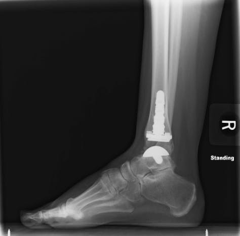
In this surgery, a round metal ball is implanted into the talus, which is an important bone in your ankle. A metal implant is also implanted into the bottom of your tibia, which is the big bone in your calf. A plastic spacer is placed between these two pieces, which allows the tibia to slide smoothly on the talus, just like it does in healthy ankles.

After surgery, a patient will typically be in a surgical boot for 8-10 weeks, and cannot put weight on that foot for 4-6 weeks (Devries, Scharer, & Sigl, 2015). Patients may be referred to physical therapy prior to, or immediately following the procedure for prehab or rehab of the ankle.

Immediately after the surgery, therapists help with gentle work to reduce swelling and pain and prevent tissues from binding down as scar tissue forms. As time passes, therapists help patients regain their strength and range of motion, restoring their ankle to full use. Healing from an ankle surgery is a long process, and requires months of physical therapy, but can be a good option when faced with debilitating ankle OA.

Did you recently have a Total Ankle Replacement surgery? We can help. Schedule your appointment today.

Fill out my online form.


SOURCES

Devries, Scharer, & Sigl. (2015). Total Ankle Arthroplasty Rehab Protocol. BayCare Clinic; Foot & Ankle Center.

Zhang, Y., & Jordan, J. M. (2010). Epidemiology of Osteoarthritis. Clinics in Geriatric Medicine, 26(3), 355–369. https://doi.org/10.1016/j.cger.2010.03.001. Retrieved from
https://www.ncbi.nlm.nih.gov/pmc/articles/PMC2920533/

Valderrabano, V., Horisberger, M., Russell, I., Dougall, H., & Hintermann, B. (2009). Etiology of Ankle Osteoarthritis. Clinical Orthopaedics and Related Research, 467(7), 1800–1806. https://doi.org/10.1007/s11999-008-0543-6

Spinal Stabilization Exercises and Their Role in Alleviating Lower Back Pain

By Boris Gilzon, PT, DPT, OCS, CHT

The Effectiveness of Spinal Stabilization Exercises for Back & Neck Pain

There is no standard approach treating chronic lower back and neck pain. Although this may be unfortunate for many patients to hear, the good news is that there are many conservative methods to alleviate pain.

While conditions like degenerative disk disease, spondylolesthesis, lumbar and cervical radiculopathy are rarely cured completely by conservative measures alone, physical therapy does offer a fair amount of pain relief in the long run.

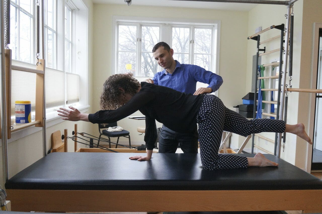
By utilizing spine stabilization exercises, our physical therapists are able to help patients reduce back and neck pain. This is an active form of treatment requiring the patient to perform exercises to strengthen the muscles and improve the stability of the spine.

Igor assisting his patient with a spine stabilization exercise.

Patients suffering from chronic spinal pain should be leery of physical therapists who mainly offer passive modalities. Examples of passive modalities include heat, electrical stimulation, and massage. Patients should be aware that passive therapeutic modalities do not have sufficient evidence to support their use in chronic spinal conditions.

Spinal stabilization exercises offer the empowerment of the patient and have plenty of research and evidence to support their effectiveness.

Pilates offers an excellent variety of spine stabilization exercise.

Extensive benefits in treating the spine of those who suffer from lower back pain have been discussed extensively in medical literature. Physical Therapists specializing in the spinal disorders are trained in recognizing the factors that affect spinal stability.

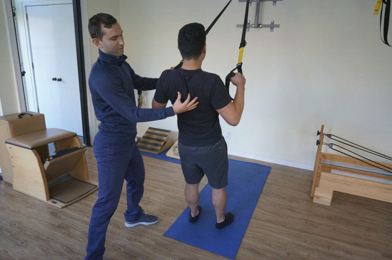
Igor Kozlov, DPT treating patient using TRX for back exercise

Components Affecting Spinal Stability

The concept of spinal stability is relatively new with research beginning during the 1970’s.

There are three components that affect spinal stability.

The first component is the passive spinal element: the bone and ligamentous structures. Studies of the cadaver spine in which the bones and ligaments are intact but the muscles were removed showed to buckle under about 20 pounds.

Spinal Ligaments - Medical Illustration Originally Sourced from Kenhub.com
Ligaments of the thoracic spine: posterior (a), anterior (b), lateral (c) and posterior with vertebral arch removed (d). 1, anterior longitudinal ligament; 2, posterior longitudinal ligament; 3, intervertebral disc; 4, ligamentum flavum; 5, intertransverse ligament; 6, supra- and interspinal ligament; 7, radiate ligament; 8, costotransverse ligament.
Originally sourced from: https://musculoskeletalkey.com/anatomy-of-the-thorax-and-abdomen/

The second component of spinal stability are the muscles that surround the spine. The muscular component provides a necessary ‘stiffening” of the spinal segment. In a healthy spine, a very modest level of muscular activity can create a sufficiently stable joint. In a degenerative disk disease, for example, there is more demand on the surrounding musculature. More strength and endurance reserve is needed to overcome an injury and pain.

Deep Muscles of the Back
Deep Muscles of the Back. Medical Illustration originally sourced from: https://pulpbits.net/7-deep-muscles-of-back-anatomy/the-deeper-muscles-of-the-back/

The third component of spinal stability are the neural elements: the central nervous system and peripheral nerves. They are akin to an orchestra conductor, coordinating the performance of various muscles, making sure they are firing at the right time, at the right amount of force.

Spinal Cord Nerves Originally Sourced from Health Jade
Spinal Cord and Nerves – Medical Illustrations originally sourced from https://healthjade.com/spinal-cord/

Multiple studies have shown patients with lower back pain make a “repositioning error” in which their spine would resume to its original position causing pain after performing a certain movement more than patients with a healthy, stabilized spine.

In physical therapy language, we call it a poor postural control.

Specific physical therapy exercises and treatment has shown effectiveness in treating chronic spinal pain.

Lumbar stabilization exercises improve muscular function which can, in turn, compensate for the structural damage to the spinal segment. A thorough dynamic assessment of the spine helps identify postural deficits.

A thoughtful exercise program is designed for each individual by the physical therapist based on their initial testing and evaluation. The most tangible benefit of a lumbar stabilization is that it gives a patient the tools to control their pain.

Interventional Pain Management

Going beyond the scope of physical therapy, interventional pain management is another passive option for chronic spinal pain. This approach serves as a temporary source of relief for patients dealing with low or medium levels of lower back pain. These techniques include performing procedures directly at the level of your dysfunction.

A pain management physician gains access to the areas causing lower back or neck pain by penetrating the surface of the skin. There is a plethora of interventional pain management options for the diagnosis and treatment of the spinal pain.

Epidural steroids are the most common example of the interventional spine management. However, the accuracy and effectiveness of interventional methods in managing lower back pain are not always clear.

In the comprehensive review article published in Pain Physician, 2013 Apr:16, the authors conducted a systematic review of literature in order to collect evidence for the effectiveness of various interventional pain management techniques in the treatment of chronic spinal pain.

The author came to the conclusion that the evidence was fair to good in 52% of therapeutic interventions. The evidence for diagnostic value fared slightly better at 62%.

One significant drawback of all passive techniques is that they do not require a participation of the patient. Without an active engagement of the patient, there is a limited self-control and independence in managing their own condition.

Do you suffer from chronic neck or back pain? Our therapists can help. Schedule your appointment today.

Fill out my online form.

Treating Hip Pain for Brooklyn Athletes

Written by Boris Gilzon, PT, DPT, OCS, CHT & Alex Ariza

Physical Therapists play an essential role in the nonsurgical treatment of hip pain. Two of the most common conditions associated with hip pain are labral tears and hip impingement.

Labral Tears

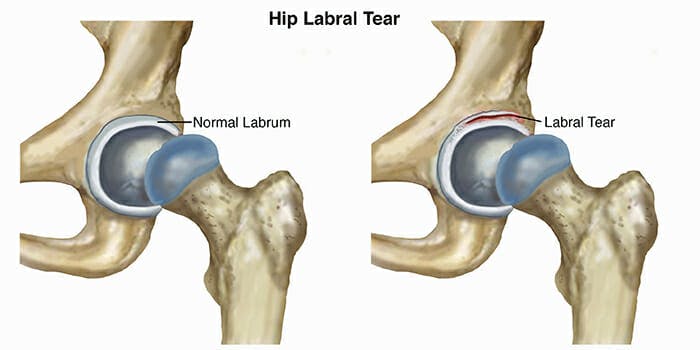
Labral tears occur when the labrum, which is the cartilage that comes between the head of the femur – your thigh bone – and your pelvis – your hip bone, begins to wear down or experiences trauma. The most common cause of labral tears is repetitive stress on the hip joint, especially for athletes participating in running and skating sports.

Labral Tear - Hip Pain
Original Image Source:
https://www.moveforwardpt.com/symptomsconditionsdetail.aspx?cid=fabdfb4e-5fb5-4077-b341-df5d04a93605

Hip Impingement

Hip impingement, also known as femoroacetabular impingement (FAI), occurs when extra bone grows on either or both the head of the femur or the acetabulum (the part of the pelvis where the femur meets – the “socket”). The extra bone creates an irregular fit and can start to rub against the cartilage or bones in a way that can damage the joint causing hip pain.

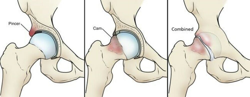
There are 2 types of hip impingement. They can occur separately or together.

Hip Impingement Types
Original image source:
https://orthoinfo.aaos.org/en/diseases–conditions/femoroacetabular-impingement/

Cam-type Impingement

The head of the femur is shaped like a sphere. It acts as the “ball” for “ball and socket” joint in your the hips. In this deformity, an excessive bone growth around the head of the femur creates an irregular shape that makes it difficult to rotate in the acetabulum, your “socket”.

The misshapen overgrowth contacts the cartilage that lines the hip socket, the labrum, and can cause it to become worn and frayed. This can lead to hip instability and pain.

Pincer-type Impingement

Pincer impingement is when the acetabulum (hip socket) protrudes around the femur head, decreasing the space of the joint. Overtime with repetitive contact, hip flexion (bending over, sitting, walking) leads to tearing and inflammation of the cartilage that envelopes and protects the joint (labrum).

Symptoms

With either one of these conditions you may experience:

  • Increased pain with prolonged sitting and leaning forward in the sitting position.
  • Sharp pain during squatting, changing direction during running, pivoting and lateral motions./li>
  • Stiffness and deep aching in the front of the hip. Occasional groin pain.
  • Decrease ability to turn your thigh inward.

Treating Hip Pain with Physical Therapy

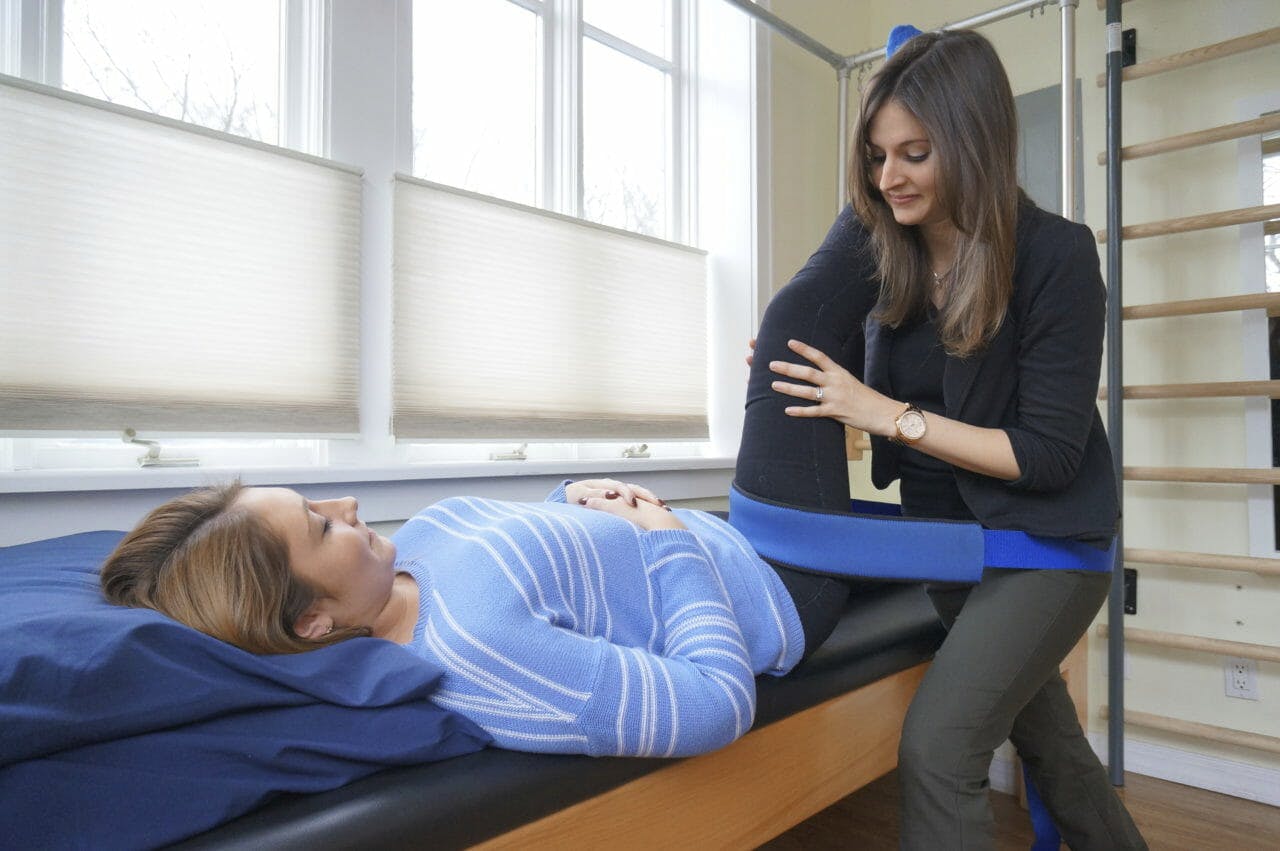
Kristin Treating Patient - Hip Mobility 2 | Park Sports Physical Therapy

Treatment of hip pain starts with a comprehensive evaluation of movement of your hip and surrounding joints.

Our therapists will test the strength of the muscles around your hip and observe your movements to properly diagnose the condition. During each session, our therapists will instruct and educate you about your body mechanics, the origin of your injury, and exercises you can perform to prevent future injury to the hip and strengthen the muscles around the hip and other parts of your body to stabilize and correct any imbalances.

Your orthopedic doctor may order X-rays and an MRI to confirm the diagnosis.

Hip impingement and labral tears may be concurrent with lower back pain and pelvic floor dysfunction.

Once your weaknesses and imbalances are identified, the goal of therapy is to restore normal force coupling and to develop strategies to compensate if your condition is chronic.

Strengthening of weak muscles and stretching tight muscles can help to avoid the progression of hip impingement. Manual therapy techniques may also be employed restore flexibility of the joint.

At Park Sports Physical Therapy, one of our therapists, Svetlana Lazarev, PT, is an advance hip clinician certified by the Hospital for Special Surgery (HSS).

Hip Arthroscopy Post Operative Rehabilitation

If you do end up having surgery for your hip pain, our therapists follow an established post-surgical protocol. Physical therapy is known to shorten recovery time and ensures that your body is healing properly. Our therapists help to promote safe healing by limiting the weight you put on the operated leg, exercises, stretching, and manual therapy.

The next stage of postoperative rehabilitation focuses on improving your range of motion and strength. During your treatment, our physical therapists can keep your doctor and orthopedic surgeon informed of your progress if you choose.

After 4 to 6 months of treatment following surgery, you’ll be able to return to your normal activities.

Do you currently suffer from hip pain? We can help.

Fill out my online form.