The Schroth Method for Scoliosis

 
The Schroth Method for scoliosis utilizes a three dimensional approach to elongate the trunk and correct imbalances of the spine. The goal is to develop the inner muscles of the rib cage in order to change the shape of the upper trunk and to correct any spinal abnormalities.

Developed by Katharina Schroth

The Schroth Method was originally developed by Katharina Schroth in Germany. The method uses customized exercises to return the body’s posture to a more natural position. It is standard treatment for scoliosis in many European countries, and is gaining trust and popularity in the United States.
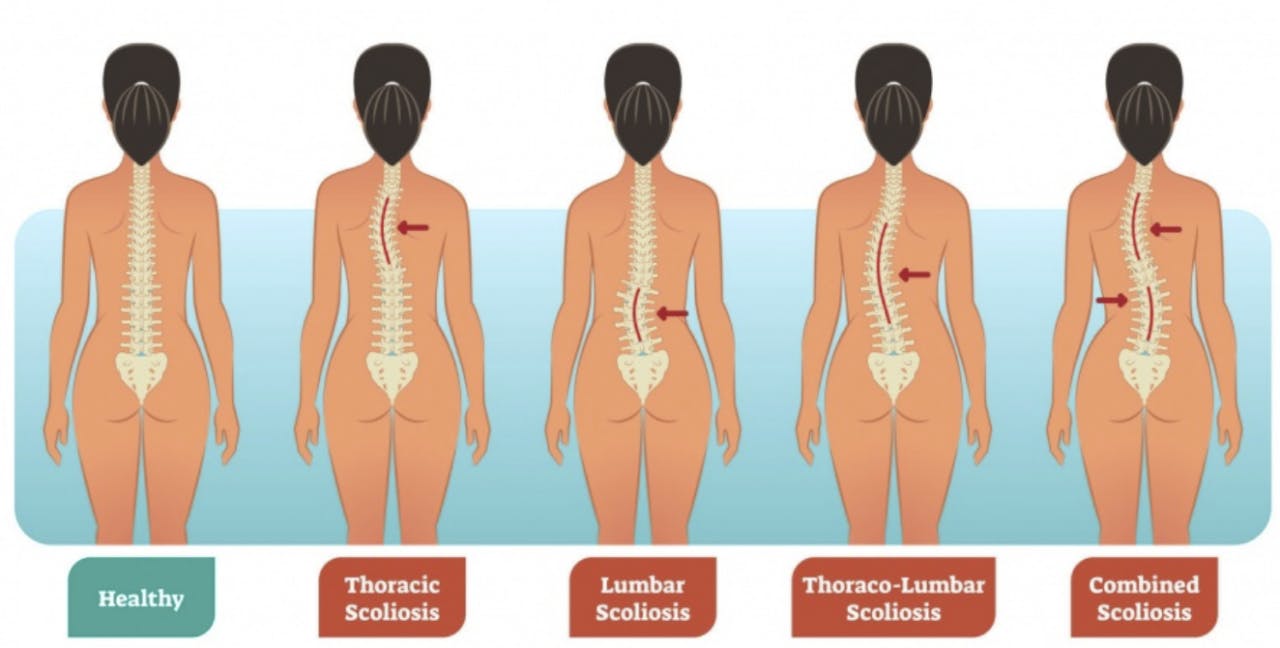
Scoliosis Symptoms

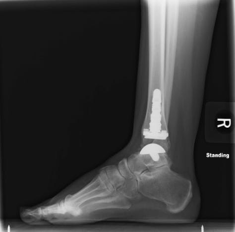
Symptoms of Scoliosis vary. Pain does not always accompany scoliosis. As a result, scoliosis can present in a variety ways such as abnormal trunk lean, uneven rib cage/shoulders or even back pain. If you suspect scoliosis contact your primary care provider. Prior to Schroth treatment, an x-ray is needed as scoliosis can present differently externally due to overlying musculature and does not give us the full picture. So, knowing the bony anatomy allows us to monitor your progress and tailor your treatment to your specific curvature.

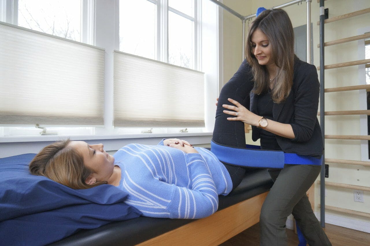
Treatment at Park Sports PT

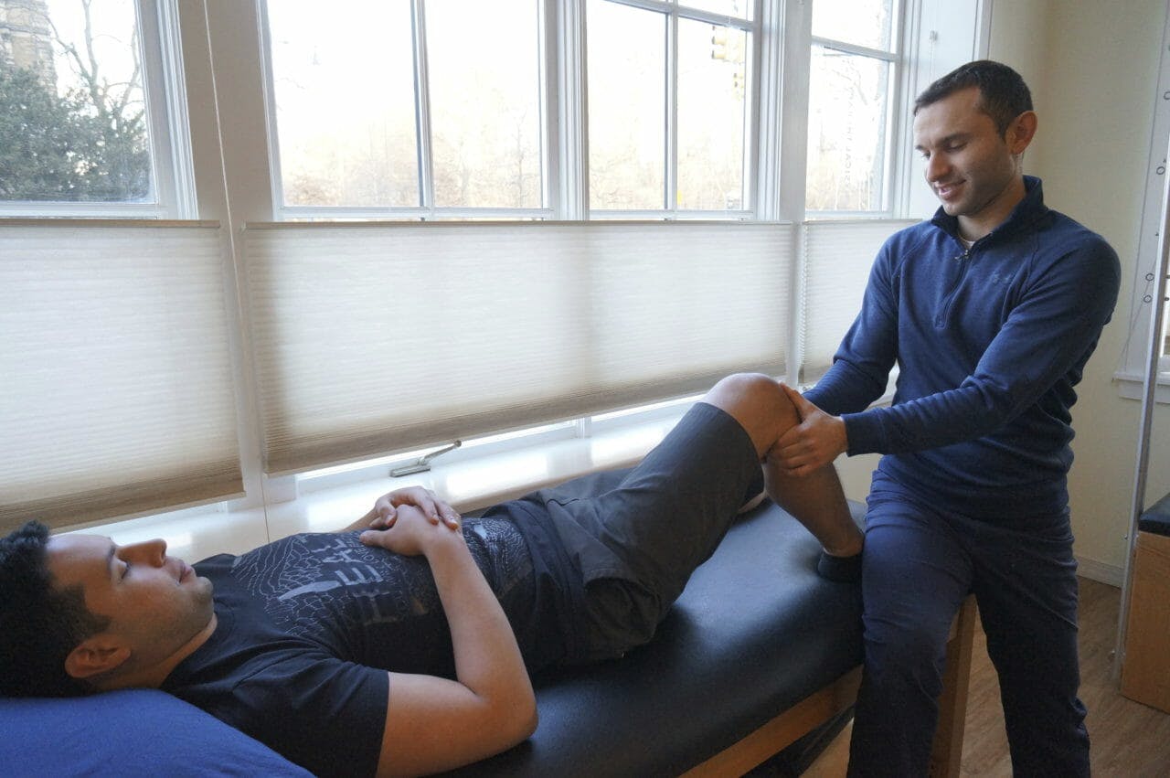
Getting treatment at Park Sports PT means you are working with certified therapists trained in the Schroth Method. They are partners in your health and wellness. Your therapist will create a personalized plan just for you and your specific condition. Our treatment approach can treat scoliosis patients of all ages. It can also can be utilized to treat in all stages of scoliosis, including after surgery.

Throughout treatment, we look closely at the three-dimensional curve in the spine. We teach patients very specific ways to correct that curve or scoliosis posture. Once patients have recognized their corrected posture, we teach them breathing and muscle activation techniques to hold that correction. Essentially, we help train the motor neurons to sense when they’re in a corrected posture versus when they’re in their scoliosis posture.

The Schroth Method takes a lot of repetition and commitment from the patient to learn these techniques. That’s why home exercises are also recommend at least five days a week for a half hour each time.

Trial results show improved patient outcomes

Several trials have found that physical therapy scoliosis-specific exercises lead to improved patient outcomes. This includes less pain and improved muscular strength, muscular endurance and self-image. In one study, spinal deformity improved in 69 percent of patients who completed Schroth exercises. This compared to only 6 percent in patients who did not complete their exercises. The Schroth Method at Park Sports PT works.

Your Park Sports PT team will create a personalized plan and will help prepare you to work on your strength and posture at home. The result is long term care and a stronger and healthier body.

Contact Us

If you or your child has any symptoms or have visited your primary care provider and are looking for treatment, contact us today to learn more. We are here to help.

Fill out my online form.


Brooklyn Half Marathon Crash Course Recap

We had a blast last night hosting the Crown Heights Running Club at our Clinton Hill location for our Brooklyn Half Marathon Crash Course.

The presenters included physical therapist and owner of Park Sports Physical Therapy, Boris Gilzon, PT, DPT, OCS, CHT, Certified Coach for the Road Runner Clubs of America, Nate Turner, and nutritionist, Tara Mardigan, MS, MPH, RD, AKA “The Plate Coach.”

They shared a lot of great information with us. This post will serve as a brief recap of some of the material that was reviewed.

Dynamic Stretching VS Static Stretching

Dynamic stretching is preferred over static stretching. You will want to focus on “warming up” your muscles before a run or undertaking any form of exercise. This can be in the form of jumping jacks, lunges, or any other full body movement. Core exercises are strongly encouraged.

There is no correlation between stretching and preventing injury, but stretching is still very beneficial in other ways. Boris recommends holding a stretch for twenty seconds or more for the muscles to get the full benefit of the stretch.

Tara recommends staying properly hydrated as that also plays a role in the performance and flexibility of your muscles.

Anywhere from 7-10 minutes of dynamic stretching will be enough to get you warmed up.

Cross Training

Cross training is useful, but depending on your goals, whether they be increasing mileage, speed, or endurance, nothing can replace running.

Boris and Nate both recommend strategic planning in terms of setting up a schedule for training to achieve your goals. The example given was a six-month training schedule. Boris recommends starting off with a light workout and running schedule at the very beginning and then increasing the intensity of theworkoutss as time progresses. This allows the body to adapt.

According to Nate, you should plan ahead and find ways to stimulate the climate of the actual race. For instance, if you are training during the colder seasons for a race that takes place during warmer seasons, you should try running in warmer temperatures some days. This could be done on a treadmill indoors with higher heat. Don’t forget to stay hydrated during these experiments!

Identifying Pre-Existing Structural Issues to Avoid Injury

Having a pre-existing injury or structural issue can lead to more serious injuries down the line. Both Nate and Boris strongly advise against working through the pain during training. Structural issues can be evaluated by a medical professional, physical therapist, or even an athletic trainer trained in identifying imbalances in the body.

A physical therapist will be able to assess your body’s strengths and weaknesses and will be able to offer valuable insight as to how you can improve your odds to avoiding injury, whether they be through strengthening exercises, modifying certain movements, correcting postural issues, or stretching and manual therapy.

If you are interested in getting a movement evaluation done by one of our physical therapists, schedule your appointment here.

Increasing Mileage Safely

Boris and Nate mentioned when training for a half marathon or even a full marathon, it’s best to work your way up to running that distance.

Instead of flat-out running thirteen miles, you could distribute a certain amount of miles each day that adds up to the full thirteen miles.

For example, on Monday you run four miles, Wednesday you run four miles, and Friday you run five miles. You can slowly increase your mileage safely in this manner instead of just deciding to run ten miles one day.

Nutrition for Runners

Tara put together an excellent handout reviewing some excellent advice in terms of nutrition, rest periods, and more. Here’s a PDF of the handout for those of you who couldn’t make it last night.

Wrapping Up

To those of you running the Brooklyn Half Marathon, we wish you the best of luck. You have trained hard and whether you aim to break a new personal record, finish the race, or have a set time that you would like to finish, we are here to help.

Want to learn more about Park Sports Physical Therapy and get started? Fill out this form here.

AlterG Anti-Gravity Treadmill

We are offering a 20% discount to Crown Heights Running Club’s members interested in trying out the AlterG Anti-Gravity Treadmill.

Schedule your first run today! Call 718.230.1180

Learn more about the AlterG treadmill and our rates here.

What Is A Total Ankle Replacement Surgery?

By: Allison Benson, Physical Therapy student at Hunter College, graduating in May 2018
Worked with Kristin Romeo, PT, DPT

With injury and with age, the joints of your body can be damaged by osteoarthritis, causing painful, aching joints. This pain can follow you throughout the day. You may feel stiff waking up, feel a dull ache when taking your dog for a walk, or feel a painful grinding as you stand up from sitting or as you climb the stairs.

Osteoarthritis (OA) is the most common joint disorder in the United States and is more common in women than men, according to an article published by Zhang & Jordan (2010). In healthy joints, where two or more bones meet and rub together, the bone surfaces are covered by a slippery substance called hyaline cartilage. This cartilage helps make your joints move smoothly and painlessly. With OA, this cartilage has broken down, leaving the bones exposed to each other, creating a grating or “bone on bone” feeling.

When a joint with OA becomes very painful, surgeons often recommend a total joint replacement—you probably know someone who has had a total knee or total hip replacement due to OA. Hips and knees are common sites for OA to develop, both because they move a lot, and because they carry the weight of the body.

You may not have heard of a total ankle replacement, though. Although ankles are also weight-bearing and mobile, they develop OA much less common; only about 1% of the population develops ankle OA (Valderrabano et al., 2009). This means many fewer people have ankle surgeries related to OA.

Another reason you may not have heard about ankle replacement is that it was a relatively unpopular surgery until recently. Total ankle replacements are complicated because there are a lot of important structures packed into a small area at the ankle. They also were associated with a very high failure rate, with surgeons needing to go back in and complete additional surgeries to replace, remove, or adjust the hardware they had placed.

That said, the popularity and success rate of total ankle replacements are on the rise.

In this surgery, a round metal ball is implanted into the talus, which is an important bone in your ankle. A metal implant is also implanted into the bottom of your tibia, which is the big bone in your calf. A plastic spacer is placed between these two pieces, which allows the tibia to slide smoothly on the talus, just like it does in healthy ankles.

After surgery, a patient will typically be in a surgical boot for 8-10 weeks, and cannot put weight on that foot for 4-6 weeks (Devries, Scharer, & Sigl, 2015). Patients may be referred to physical therapy prior to, or immediately following the procedure for prehab or rehab of the ankle.

Immediately after the surgery, therapists help with gentle work to reduce swelling and pain and prevent tissues from binding down as scar tissue forms. As time passes, therapists help patients regain their strength and range of motion, restoring their ankle to full use. Healing from an ankle surgery is a long process, and requires months of physical therapy, but can be a good option when faced with debilitating ankle OA.

Did you recently have a Total Ankle Replacement surgery? We can help. Schedule your appointment today.

Fill out my online form.


SOURCES

Devries, Scharer, & Sigl. (2015). Total Ankle Arthroplasty Rehab Protocol. BayCare Clinic; Foot & Ankle Center.

Zhang, Y., & Jordan, J. M. (2010). Epidemiology of Osteoarthritis. Clinics in Geriatric Medicine, 26(3), 355–369. https://doi.org/10.1016/j.cger.2010.03.001. Retrieved from
https://www.ncbi.nlm.nih.gov/pmc/articles/PMC2920533/

Valderrabano, V., Horisberger, M., Russell, I., Dougall, H., & Hintermann, B. (2009). Etiology of Ankle Osteoarthritis. Clinical Orthopaedics and Related Research, 467(7), 1800–1806. https://doi.org/10.1007/s11999-008-0543-6

How Physical Therapy Works To Eliminate Knee Pain

By Boris Gilzon, PT, DPT, OCS, CHT

In a 2006 health survey conducted by the National Health Interview Survey (NHIS), knee pain was reported as the second most common cause of chronic pain in America.

Another surprising statistic comes from the Society for Academic Emergency Medicine. They reported that “the knee is the most commonly injured joint by adolescent athletes with an estimated 2.5 million sports-related injuries presenting to [Emergency Departments] annually.”

Some studies even show us that there has been an increase in the amount of knee replacement procedures over the last few years. Researchers say this is caused by two major factors: the first being the obesity epidemic and the second being that we are living longer lives. While living longer is great, it also puts more years of wear and tear on our bodies which can lead to osteoarthritis.

So what can you do to prevent knee pain or if you already suffer from knee pain, how can you better manage it and get out of pain?

The knee joint can only move in one plane, like a door hinge, and does not accommodate well to external stress that falls outside of its natural axis. For example, imagine being pushed from the side while your feet are firmly planted. This is the most common mechanism leading to a knee injury. Anterior Cruciate Ligament (ACL) injuries, as well as meniscus tears, normally occur this way.

The knee joints bear multiples of your body weight in running and jumping. Climbing up the stairs, for example, loads your knee joint 2.5 times your body weight.

The knee is considered a biomechanical link between the hip and the ankle/foot complex. Dysfunction in any of these joints can negatively affect the others in the chain. Repeated abnormal stress can take a toll on the knee joint.

Knee pain is one of the most common conditions our therapists treat in our clinics. Our therapists know how to take care of a variety of injuries and conditions for people of all ages. Early intervention of knee pain will improve your quality of life, mobility, and prevent loss of muscle strength and instability.

Types of Knee Injuries

There are two categories that a knee injury can fall into: 1) acute/traumatic and 2) chronic/repetitive stress. Acute injuries are when the incidents occur immediately, like a fall, car accident, landing in a strange way, twisting/pivoting quickly, etc. Many sports injuries, especially sprains and strains, fall under this category.

Chronic injuries are caused by repetitive stress over a long period time. Poor posture and/or body mechanics can play a major role in chronic conditions. Physical therapy can be very beneficial in correcting these issues.

Osteoarthritis

osteoarthritis knee joint
Osteoarthritis of the knee joint. Medical illustration original source: https://drcolinmacleod.com/platelet-rich-plasma-arthritis

Knee pain can be caused by degenerative changes in osteoarthritis. Arthritis is when the cartilage cushioning the bones wear down leading to swelling, stiffness, and pain.

Unfortunately, in the cases of the knee pain due to severe osteoarthritis, Physical therapy intervention is limited and one should consult with an orthopedic doctor to assess whether a total knee replacement is appropriate.

Knee pain is more commonly seen in people who do not yet have visible arthritic changes on radiographic examination. Those people are engaged in various physical activities while struggling with the knee pain during and after the activity.

Anterior Knee Pain aka Runner’s Knee

knee medical illustration patella
The patella “floats” between the thighbone and shinbone in the trochlear groove. Medical illustration original source: https://orthoinfo.aaos.org/en/diseases–conditions/patellofemoral-arthritis/

Anterior knee pain or the “Runner’s Knee” is related to an abnormal motion of the kneecap in the trochlear groove. It causes an irritation and eventual wearing out of the cartilage on the back of your kneecap. The knee pain gets worse when you first stand up, run and going downstairs. The knee pain worsens while performing your physical activity.

Patellar Tendonitis aka Jumper’s Knee

patellar tendonitis
Patellar Tendonitis Medical Illustration Original Source: https://www.vivehealth.com/blogs/resources/patellar-tendonitis

Patellar tendonitis, also known as “Jumper’s Knee,” is another activity related condition that is caused by repetitive motion. The knee pain, in this case, originates in the patellar tendon. A structure that connects your quadriceps muscle to the lower leg through the kneecap. When your quadriceps muscle is overloaded it causes an inflammation of the tendon, thus contributing to the knee pain. The symptoms are usually more pronounced when you are at rest and when you initiate your activity. In more severe and chronic cases the knee pain prevents you from participating in sports.

Knee Pain Rehabilitation and Treatment

The key in the rehabilitation of the knee pain is a correct biomechanical analysis of your kinetic chain. An exercise regimen performed at the proper angles and positions. Prescribed activities help to avoid further irritation of the joint and yet strengthening the weak elements. If you suffer from the knee pain, it does not mean that you need to halt your physical activities. Physical Therapist at Park Sports have the tools and knowledge to get you ‘back in the game”.

Do you currently suffer from knee pain? We can help. Get started by filling out the form below.

Fill out my online form.


Now Offering Pay-As-You-Go Rates for the AlterG Antigravity Treadmill in Clinton Hill

Sean “P. Diddy” Combs using the AlterG AntiGravity Treadmill
Twitter post of Sean “P. Diddy” Combs using the AlterG AntiGravity Treadmill

The Perfect Solution for Recovering Athletes

If you’re a Brooklyn athlete and recently sustained an injury or underwent surgery on your lower body we have the perfect solution to get you running sooner. The AlterG Anti Gravity Treadmill uses NASA’s patented Differential Air Pressure Technology to “unweight” you, making it possible to run at a fraction of your body weight. You’ll be able to improve your aerobic conditioning and put less stress on your joints – hips, knees, and ankles – while maintaining strength and endurance.

Not Just for Athletes

The AlterG can also benefit:

  • Senior Citizens looking to stay active
  • People with obesity looking workout and lose weight
  • Anyone with past injuries or arthritis looking to workout with less impact and stress on their joints

Rates & Monthly Memberships

Your first 30-minute trial run is only $20.00 (normally $25.00). Call 718.230.1180 to schedule your first run!


Time Price
Under 30 Minutes  $1.00/minute
30 Minutes $25.00
30 Minutes – Three Scheduled Sessions $50.00
Monthly Memberships Price
Up to 45 minutes – Three Scheduled Sessions Per Week $100.00
Up to 90 minutes – Three Scheduled Sessions Per Week $200.00

Want more information? Fill out the form below.

Fill out my online form.

Frozen Shoulder Treatment – Regaining Mobility Through Physical Therapy

Written by Edward Umheiser, PT, DPT
Edited by Alex Ariza

Injuries to the shoulder are fairly common in everyday life. Strains and sprains resulting from repetitive activities such as throwing a ball or twisting your arm while reaching behind your car seat happen all the time, and usually heal on their own in only a few days. But what happens if your shoulder suddenly starts to lose its normal range of motion for an unexplained reason?

In this article, I would like to talk about the diagnosis known as adhesive capsulitis, more commonly referred to as frozen shoulder.

What is Frozen Shoulder?

Frozen shoulder is a fairly common diagnosis, but is not well understood and does not always have a known cause. Many people develop frozen shoulder following a surgical procedure to the shoulder, or after an injury that results in the shoulder needing to be immobilized for a short period of time. However, in some cases, some people develop symptoms with no known trigger.

The symptoms are fairly clear-cut – a marked loss of normal range of motion and stiffness of the shoulder joint that may or may not be accompanied by pain.

This shoulder stiffness affects motion in all directions such as reaching up in the air or trying to place the hand behind the back or behind the head. Adhesive capsulitis begins as an inflammatory process within the shoulder and leads to scar tissue formation which can restrict shoulder motion.

For some people, this scar tissue formation can lead to pain in the shoulder joint, especially with movement and during the evening hours while trying to sleep.

The Stages of Frozen Shoulder

Physical Therapist, Edward Umheiser, DPT treating patient with frozen shoulder.
Physical Therapist, Edward Umheiser, DPT treating a patient with frozen shoulder.

There are several stages of frozen shoulder each with their own unique characteristics:

Inflammatory Stage

The inflammatory stage lasts approximately three months and is characterized by pain with shoulder movement in most directions. Pain is often sharp with movement at the end of the range, and there is an ache at rest. Pain is also commonly felt at night, making it difficult to fall asleep.  The range of motion may still be normal at this early stage.

Freezing Stage

Edward Umheiser, DPT measures the shoulder's range of motion.
Edward Umheiser, DPT measures the shoulder’s range of motion.

The freezing stage typically lasts 3-9 months. This is when flexibility of the shoulder begins to reduce due to pain. At this time, people often begin to notice difficulty reaching overhead, or reaching behind their back. This stage, known as the frozen stage, is when the stiffness is most severe. The inflammatory process inside the shoulder joint is starting to decrease at this stage so pain is lessened or non-existent. Over the past several months a thick layer of scar tissue has formed around the shoulder capsule, which makes it difficult to move. This is the stage when most people are diagnosed with frozen shoulder.

Thawing Stage

Edward Umheiser, DPT treating a patient with frozen shoulder. With consistent physical therapy sessions, a patient can begin seeing an increased range of motion.

Finally, the thawing stage is when shoulder motion gradually starts to return to normal. This stage can last anywhere from 9 months to two years and may leave some minor limitations in shoulder range of motion afterward.

Treating A Frozen Shoulder

Edward Umheiser, DPT teaching the patient shoulder exercises.

Physical therapy along with treatment provided by your doctor such as cortisone injections are the first line of defense in reducing the time a patient may experience the symptoms related to a frozen shoulder.

A doctor will typically take some images of the shoulder to rule out other possible structural involvement such as the rotator cuff or the labrum.

At that point, physical therapy treatment 2-3x a week is generally recommended where manual shoulder stretching, massage, mobilization, and exercises are performed to keep the shoulder as pain-free as possible, and to help increase the range of motion.

It is also important to continue to perform the prescribed stretching exercises at home under the guidance of a physical therapist. Generally, the time it takes for a frozen shoulder to “thaw” can be halved under physical therapy treatment. Most patients do not need surgical intervention to correct this diagnosis.

Steps to Take If You Suspect Having a Frozen Shoulder

If you have noticed that you have a lot more difficulty with overhead shoulder motion following a shoulder injury, let your doctor know. If this has persisted for several months, it may be a sign of adhesive capsulitis. An orthopedist can help you make that diagnosis, and physical therapy can help you get onto the road to recovery.

Schedule Your Physical Therapy Appointment Today To Begin Frozen Shoulder Treatment

Fill out my online form.

Treating Hip Pain for Brooklyn Athletes

Written by Boris Gilzon, PT, DPT, OCS, CHT & Alex Ariza

Physical Therapists play an essential role in the nonsurgical treatment of hip pain. Two of the most common conditions associated with hip pain are labral tears and hip impingement.

Labral Tears

Labral tears occur when the labrum, which is the cartilage that comes between the head of the femur – your thigh bone – and your pelvis – your hip bone, begins to wear down or experiences trauma. The most common cause of labral tears is repetitive stress on the hip joint, especially for athletes participating in running and skating sports.

Labral Tear - Hip Pain
Original Image Source:
https://www.moveforwardpt.com/symptomsconditionsdetail.aspx?cid=fabdfb4e-5fb5-4077-b341-df5d04a93605

Hip Impingement

Hip impingement, also known as femoroacetabular impingement (FAI), occurs when extra bone grows on either or both the head of the femur or the acetabulum (the part of the pelvis where the femur meets – the “socket”). The extra bone creates an irregular fit and can start to rub against the cartilage or bones in a way that can damage the joint causing hip pain.

There are 2 types of hip impingement. They can occur separately or together.

Hip Impingement Types
Original image source:
https://orthoinfo.aaos.org/en/diseases–conditions/femoroacetabular-impingement/

Cam-type Impingement

The head of the femur is shaped like a sphere. It acts as the “ball” for “ball and socket” joint in your the hips. In this deformity, an excessive bone growth around the head of the femur creates an irregular shape that makes it difficult to rotate in the acetabulum, your “socket”.

The misshapen overgrowth contacts the cartilage that lines the hip socket, the labrum, and can cause it to become worn and frayed. This can lead to hip instability and pain.

Pincer-type Impingement

Pincer impingement is when the acetabulum (hip socket) protrudes around the femur head, decreasing the space of the joint. Overtime with repetitive contact, hip flexion (bending over, sitting, walking) leads to tearing and inflammation of the cartilage that envelopes and protects the joint (labrum).

Symptoms

With either one of these conditions you may experience:

  • Increased pain with prolonged sitting and leaning forward in the sitting position.
  • Sharp pain during squatting, changing direction during running, pivoting and lateral motions./li>
  • Stiffness and deep aching in the front of the hip. Occasional groin pain.
  • Decrease ability to turn your thigh inward.

Treating Hip Pain with Physical Therapy

Kristin Treating Patient - Hip Mobility 2 | Park Sports Physical Therapy

Treatment of hip pain starts with a comprehensive evaluation of movement of your hip and surrounding joints.

Our therapists will test the strength of the muscles around your hip and observe your movements to properly diagnose the condition. During each session, our therapists will instruct and educate you about your body mechanics, the origin of your injury, and exercises you can perform to prevent future injury to the hip and strengthen the muscles around the hip and other parts of your body to stabilize and correct any imbalances.

Your orthopedic doctor may order X-rays and an MRI to confirm the diagnosis.

Hip impingement and labral tears may be concurrent with lower back pain and pelvic floor dysfunction.

Once your weaknesses and imbalances are identified, the goal of therapy is to restore normal force coupling and to develop strategies to compensate if your condition is chronic.

Strengthening of weak muscles and stretching tight muscles can help to avoid the progression of hip impingement. Manual therapy techniques may also be employed restore flexibility of the joint.

At Park Sports Physical Therapy, one of our therapists, Svetlana Lazarev, PT, is an advance hip clinician certified by the Hospital for Special Surgery (HSS).

Hip Arthroscopy Post Operative Rehabilitation

If you do end up having surgery for your hip pain, our therapists follow an established post-surgical protocol. Physical therapy is known to shorten recovery time and ensures that your body is healing properly. Our therapists help to promote safe healing by limiting the weight you put on the operated leg, exercises, stretching, and manual therapy.

The next stage of postoperative rehabilitation focuses on improving your range of motion and strength. During your treatment, our physical therapists can keep your doctor and orthopedic surgeon informed of your progress if you choose.

After 4 to 6 months of treatment following surgery, you’ll be able to return to your normal activities.

Do you currently suffer from hip pain? We can help.

Fill out my online form.

What We Can Learn From Angels Pitcher Shohei Ohtani’s Elbow Injury

Written by Boris Gilzon, PT, DPT, OCS, CHT and Alex Ariza

If you’re a fan of baseball you may have heard that New Los Angeles Angels pitcher, Shohei Ohtani, has a damaged Ulnar Collateral Ligament (UCL) in his pitching arm.

This is bad news for Ohtani, as his career relies heavily on the function his elbow. Although reports show that Ohtani only has a first-degree sprain, consistent use of his elbow can present serious problems down the line.

In the world of physical therapy, elbow pain does not get enough exposure and is often ignored. More often than not, people continue performing their daily activities that can exacerbate injury to the elbow and cause damage to the joint and ligaments.

Ulnar Collateral Ligament (UCL) Injury

As Physical Therapists, we see sports injuries in a different light, especially when they make the news. This post will share some of the intimate knowledge we possess from years of treating a number of injuries and conditions that athletes may face.

The Ulnar Collateral Ligament, located in the elbow, is a complex structure consisting of three bands. It is challenged more when the elbow is in flexion. During full elbow extension, the bony congruity provides additional stability.

Ulnar Collateral Ligament Anatomy

Ulnar Collateral Ligament injury is common in many overhead sports. The forces that can lead to injury on the elbow are generated when the elbow goes from flexion to extension at a high velocity. In Ohtani’s case, this would be frequently pitching at a consistent speed of 100 mph.

This injury occurs from repetitive valgus (a condition in which the bone segment distal to a joint is angled outward) stress on the medial (inner aspect) of the elbow. This kind of elbow sprain commonly occurs in baseball pitchers. It is less frequent in racket sports, volleyball, and hockey, but can still occur in athletes playing those sports.

Damage to the ligament can be caused by sudden trauma or a gradual stress. The most typical sign is a pain in the inner elbow while performing a physical activity. Patients commonly describe an elbow sprain as a twinge or sharp pain when pushing up, such as getting up from the chair.

If a ligament is compromised a patient may develop:

  • A sense of looseness or instability in the elbow.
  • Irritation of the ulnar nerve (aka the “funny bone”): This is felt as a tingling sensation or numbness in the small finger and ring finger.
  • Decreased ability to throw a baseball or other object overhead.

Treatment for Ulnar Collateral Ligament Injury

Ulnar Collateral Ligament injury can happen to anyone at any age. Parents and coaches should be aware of the issue and be alert if the child complains of the pain in the elbow.

Physical Therapy is the best first line of defense following this diagnosis. At Park Sports Physical Therapy, our Physical Therapists are skilled and experienced in treating elbow instability and ulnar collateral ligaments tears.

The treatment approach is based on thorough examination and biomechanical analysis of the activity that caused the condition.

It includes:

  • Strengthening weak muscle group
  • Activity modification.
  • Manual therapy and therapeutic modalities.

Post-Surgical Recovery

In cases of severe damage and instability, the ligament is reconstructed in what is commonly known as a Tommy Johns procedure. Physical Therapy treatment in this case follows an established post-surgical protocol. Our therapists work closely with an Orthopedic Surgeon to optimize post-surgical recovery and achieving the highest functional outcomes.

Do you suffer from Ulnar Collateral Ligament Injury? Take the first step to get out of pain by scheduling your appointment.

Fill out my online form.

Post Surgical Rehabilitation in Brooklyn

If you just had an operation for a muscle or joint problem, then you need want to experienced clinicians that provide some of the best post surgical rehabilitation in Brooklyn.

A Park Sports Physical Therapy treatment program will help shorten the road to recovery after surgery.

After an injury occurs, the first thing most people think about is recovering as quickly as possible and getting back to doing what they love.  For most injuries, physical therapy is considered the best possible option to help accomplish this goal.  Physical therapists are experts at diagnosing injuries and creating treatment programs that are personalized for each patient based on their particular condition.  The treatments they provide are also supported by countless research studies that continue to come out.

Sometimes You Need a Surgeon…then Great Post Operative Rehabilitation in Brooklyn

Not all injuries can be treated with physical therapy alone.  In some cases, a person’s injury might be too severe, or they may have already tried physical therapy and it failed to bring about any major improvements.  In these cases, surgery may be recommended as the next option.

Patients with severe osteoarthritis of the hip or knee, chronic low back pain, tears of the meniscus, ACL or Achilles tendon and many other injuries may consider surgery if their condition doesn’t improve trying after physical therapy.  These patients should carefully weigh the benefits and risks of surgery, since there is no guarantee that it will lead to a successful outcome.  If the patient decides to go through with surgery, it’s also important to realize that it may take some time to completely recover.

Once again, this is where our physical therapists can help.  One of the best possible ways to ensure a positive outcome and shorten the road to recovery is by participating in a physical therapy program after undergoing surgery.  Depending on the surgical procedure, physical therapy can begin right away or soon afterwards, and it will focus on bringing the patient back to full strength as quickly as possible.  Treatment usually includes a variety of modalities including:

  • Neuromuscular reeducation,
  • Post-tetanic stretching,
  • Hands-on mobilization,
  • Progressive strengthening exercises,
  • Functional training with a walking device if one is being used, and
  • Education on how to return to activities with a minimal risk for future injuries.

The benefits of having post-surgical physical therapy can be seen in the results of this recent study:

The study showed that patients who received both outpatient physical therapy and a home-exercise program improved in range of motion, the 6-minute walking test, and health-related quality of life. Patients who received outpatient physical therapy also had significantly fewer days from the date of surgery to physical therapy discharge.
Reference: https://www.ncbi.nlm.nih.gov/pubmed/27851675

Nobody wants to have surgery, but it may be necessary if your injury or condition is too severe.  If you find yourself in this predicament, the best way to ensure a fast and successful recovery is to follow a physical therapy program that’s created specifically for you.

We have four physical therapy clinics here in Brooklyn.  Click here for contact information.